


A new study reveals how the brain's internal navigation system, known as grid cells, is connected to eye movements and plays an unexpected role in how we form memories of visual scenes. The research, published in Nature Communications, shows that specific brain activity patterns related to eye movements are linked to how well a person remembers an image later.
Key Takeaways
- Researchers found a link between rapid eye movements (saccades) and 'grid-like codes' in the brain's entorhinal cortex while people viewed images.
- These grid-like codes, part of the brain's spatial mapping system, were more prominent for images that were later remembered.
- Unexpectedly, individuals with lower intensity grid-like signals demonstrated better memory performance.
- The study also identified a connection between this entorhinal activity and the frontal eye fields, a brain region that coordinates eye movement.
How the Brain Maps Visual Space
When we look at our surroundings, our eyes don't move smoothly. Instead, they make quick, sharp jumps called saccades. These movements allow us to gather information from different parts of a scene, piecing together a complete picture.
Scientists have long known that a specific part of the brain, the entorhinal cortex, contains special neurons called grid cells. These cells act like a coordinate system, helping us navigate physical space. Think of it as the brain's internal GPS.
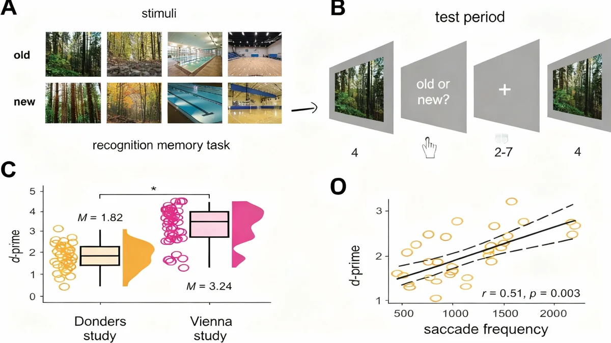
This new research explored whether this same system is used to map visual space during memory formation. The study analyzed data from two independent experiments where participants viewed a series of scene images while their brain activity and eye movements were monitored using functional magnetic resonance imaging (fMRI) and eye-tracking technology.
What Are Grid Cells?
Grid cells are neurons in the entorhinal cortex that fire at multiple locations in an environment, forming a hexagonal, grid-like pattern. This pattern provides a metric for space, allowing the brain to understand distances and directions. While discovered during studies of physical navigation in animals, researchers are now finding they play a broader role in cognition, including mapping abstract and visual information.
Connecting Eye Movements to Brain Activity
The core of the study was to see if the direction of a person's saccades correlated with the activity patterns of grid cells. Using fMRI data, researchers looked for a specific signal—a hexadirectional, or six-fold, pattern of brain activity corresponding to the direction of eye movements.
Across both datasets, a consistent finding emerged. "We consistently identified saccade-based grid-like codes in the left entorhinal cortex while participants studied scene images," the authors of the paper stated. This confirms that the brain's navigation system is active not just when we walk around, but also when our eyes explore a static image.
The analysis was specific to this six-fold pattern. When researchers tested for other patterns (like four-fold or five-fold symmetry), the effect disappeared, strengthening the evidence that they were observing the signature of grid cell activity.
An Unexpected Link to Memory Performance
The most surprising part of the research was how this brain activity related to memory. The study found that grid-like signals were present specifically for images that participants later remembered successfully. This suggests these codes are directly involved in the process of encoding memories.
However, the relationship was not what one might expect. Individuals who showed lower intensity grid-like codes actually had better recognition memory. In the Donders study dataset, there was a significant negative correlation: the weaker the grid signal, the better the participant's performance on the memory test.
Memory and Saccades
The study confirmed previous findings that more eye movements are linked to better memory. In one dataset, participants made an average of 7.95 saccades for scenes they later remembered, compared to only 6.64 for scenes they forgot. This highlights the importance of active visual exploration in forming strong memories.
Why Less Signal Might Mean Better Memory
The researchers propose a few possible explanations for this counterintuitive finding. One theory is related to cognitive efficiency. Individuals with better memory might process visual information more efficiently, requiring less neural activity in the entorhinal cortex.
"Individuals with good recognition memory might display less neural activation during memory formation, thus, encoding 'more efficiently'," the study suggests.
Another possibility is that high-performing individuals may rely more on prior knowledge or schemas to understand a scene. For example, when viewing a beach, they might activate a mental 'beach schema' which guides their eye movements more effectively, reducing the need for the brain's grid system to build a visual map from scratch.
This suggests that relying purely on the raw spatial layout of a scene (which is what grid cells map) might be a less effective strategy than integrating new information with existing knowledge.
Coordinating Brain Networks for Vision and Memory
The study also shed light on how different brain regions work together. The researchers investigated which other brain areas were active in sync with the entorhinal grid-like signals. They found a strong connection to the frontal eye fields (FEF), a region known to be critical for planning and executing eye movements.
When the grid-like signals in the entorhinal cortex were strong, activity in the left FEF was also high. This indicates a direct link between the brain's spatial map and the machinery that controls where the eyes look next.
This finding supports a model where the entorhinal cortex provides a spatial framework, which in turn informs the FEF on how to guide saccades to explore a scene. This coordinated activity helps build a coherent visual representation that can be stored as a memory.
Implications for Understanding Memory and Brain Function
This research adds to a growing body of evidence that the brain's navigation system has been adapted for functions beyond physical movement. It appears to provide a universal 'cognitive map' for organizing different types of information, including the contents of a visual scene.
The findings have several important implications:
- Understanding Memory Encoding: The study provides a neural mechanism for how visual exploration directly contributes to memory formation.
- Brain Network Dynamics: It highlights the interplay between memory regions (entorhinal cortex) and motor control regions (frontal eye fields).
- Future Research: The unexpected negative relationship between grid signal strength and memory performance opens up new questions about what constitutes 'efficient' brain processing during learning.
While direct evidence connecting single grid cell recordings to saccades in humans is still needed, this fMRI study provides a powerful, non-invasive look into this fundamental process. It reveals that every time our eyes dart across a scene, a complex coordination between our internal GPS and motor systems is at work, shaping what we see, and what we will remember.